Types of Skin Cancer
Skin cancer is the most common cancer in Australia; two in three people will be diagnosed with skin cancer in their lifetime.
When skin cancer detected early, it’s almost always completely treatable.
When skin cancer detected early, it’s almost always completely treatable.
|
Basal Cell Carcinoma (BCC)
This is the most common and usually the least serious type of skin cancer. BCCs often appear as a pearly bump, scaly patch, or a wound that doesn’t heal. They tend to grow slowly, but they can cause local tissue damage if left untreated. Treatment typically involves surgical excision, or for some mild cases, cryotherapy and topical therapies are suitable. 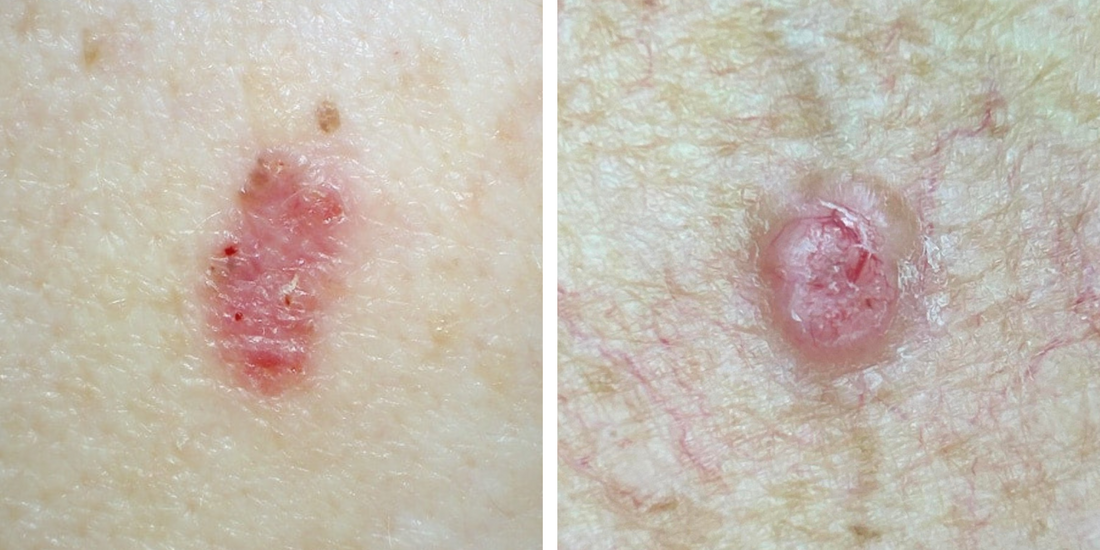
|
Squamous Cell Carcinoma (SCC)
SCCs are the second most common type of skin cancer. They often look like a crusted, scaly, or thickened patch that may bleed or feel tender. SCCs can grow more quickly than BCCs and, in rare cases, may spread to other areas of the body if not treated early. Treatment usually involves surgical excision as the most effective cure. 
|
Intraepidermal Carcinoma (IEC)
IEC is an early form of squamous cell carcinoma cancer (SCC) confined to the top layer of the skin. It often appears as a flat, red, scaly or crusted patch. IEC can progress to invasive SCC if left untreated. There are a wide range of treatment options available, including surgical excision, cryotherapy, or topical therapies. 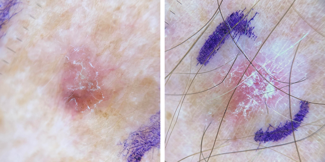
|
Melanoma
Melanoma is the least common, but most serious type of skin cancer. It can develop from an existing mole, or appear as a new spot. Melanomas can spread rapidly if not detected early, which is why regular skin checks and monitoring for new or changing spots is so important. Early melanomas can often be completely cured with surgery. 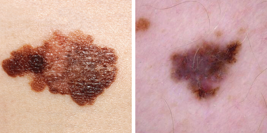
|
Common Skin Spots
Not all skin spots are dangerous. Many growths, moles, and marks on the skin are completely harmless (benign).
They may not require treatment unless they become irritated or they may be treated for cosmetic reasons.
Some spots require a biopsy if they changing in appearance, of if they look suspicious for a potential skin cancer.
They may not require treatment unless they become irritated or they may be treated for cosmetic reasons.
Some spots require a biopsy if they changing in appearance, of if they look suspicious for a potential skin cancer.
|
Seborrhoeic Keratoses
These are very common, harmless growths that often appear with age. They can appear warty, waxy, or “stuck on” the skin, and range in colour from light brown to almost black. They do not turn into skin cancer but can be removed easily if they catch on clothing or cause irritation. Sometimes they require biopsy or removal if they appear suspicious, to rule out skin cancer. 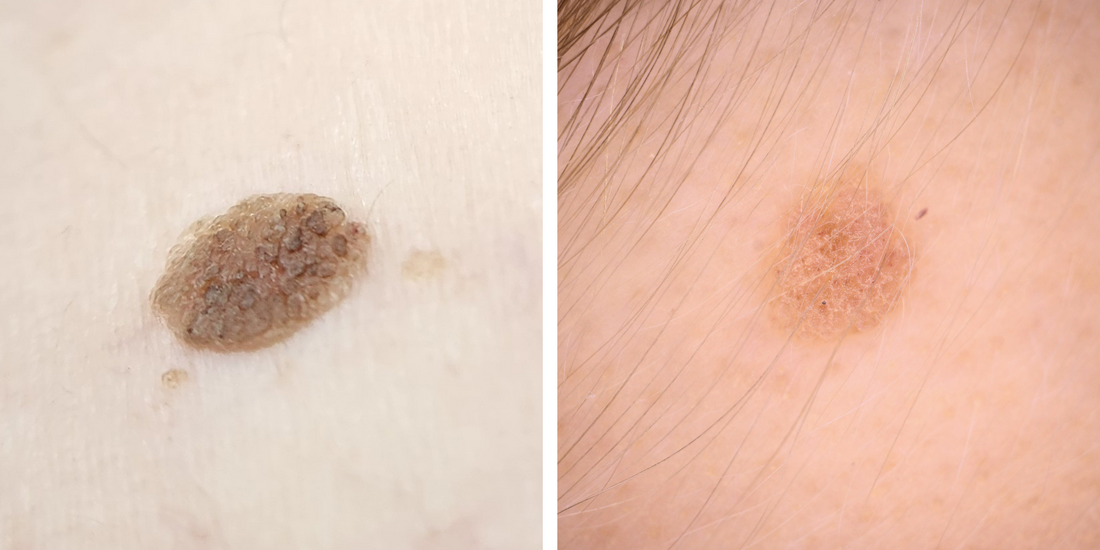
Hemangiomas
These are small, bright red or purple spots made up of tiny blood vessels. They are completely benign and very common, especially with age. They can be treated with cryotherapy, laser or surgery. 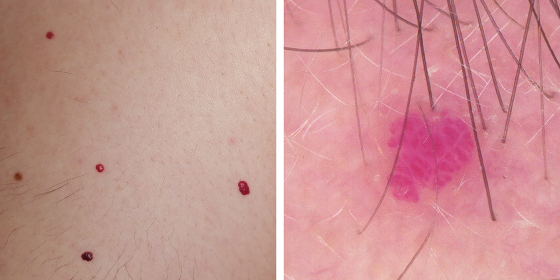
|
Solar (Actinic) Keratoses
These are rough, scaly patches that develop on sun-exposed areas such as the face, scalp, neck, and arms. They are not cancer, but can sometimes develop into a squamous cell carcinoma if left untreated. Treatment options include cryotherapy and topical treatments. Sun protection, including sunscreen (50+ SPF) is vital for prevention, and can aid in reversing this type of sun damage. 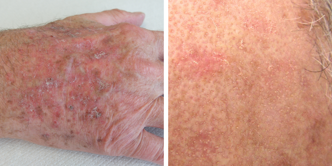
Skin Tags
Skin tags are soft, flesh-coloured growths often found on the neck, underarms, or groin. They often develop in areas under friction, and are benign. They can be treated with cryotherapy or surgery. 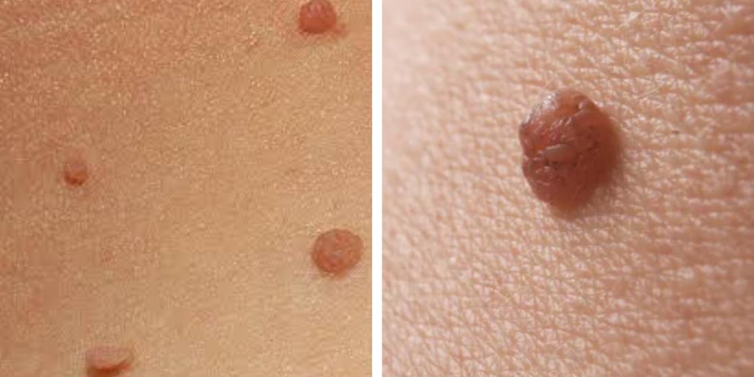
|
Moles (Naevus/Naevi)
Moles are clusters of pigment cells that are usually brown, black, or pink. They may be flat patches or raised nodules. Most moles are harmless and stable throughout the lifetime. It is important to monitor moles for changes in size, colour, or shape, as these changes may indicate a possible melanoma. Some may require biopsy or removal if they appear suspicious, to rule out skin cancer. 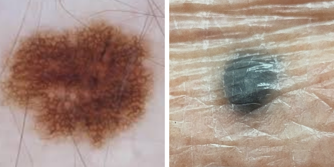
Cysts
Cysts are smooth, round lumps under the skin caused by blocked glands or trapped keratin. They’re not cancerous, though they can become inflamed or infected. If needed, they can be surgically removed. 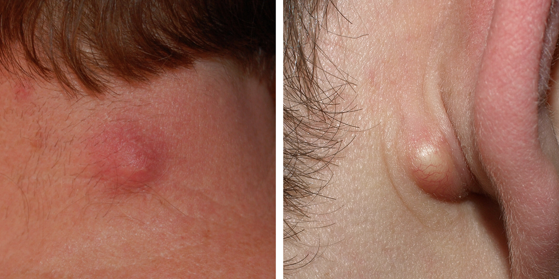
|
Your Skin Journey
Taking care of your skin is a lifelong commitment. Here’s what to expect at each step.
|
1. Skin Check
|
2. Skin Cancer Treatment
|
3. Post-treatment Care
|